
Fig. 2.
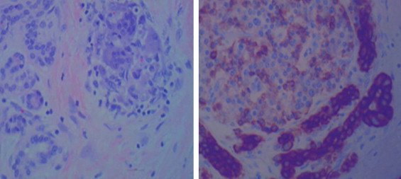
Images were taken at 400× with 75 gold scale bars. On left Hematoxylin and eosin stains images; on right are CK AE1/AE3 immunohistochemical stains. They show cancersous glands invested by inflammatory cells including osteoclast-like giant cells.