
Figure 2.
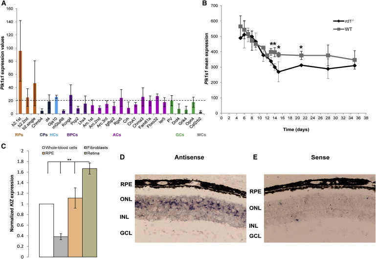
Plk1s1 Transcriptomic Analysis in the Mouse Retina
(A) Plk1s1 expression (1455558_at) in six different cell types from the mouse adult retina: rod photoreceptors (RPs), cone photoreceptors (CPs), horizontal cells (HCs), bipolar cells (BPCs), amacrine cells (ACs), ganglion cells (GCs), and microglia cells (MCs). The graph presents Plk1s1 normalized expression values. Only values higher than 20 can be considered significantly expressed. Retinal cell types were established from a library composed of 22 transgenic mouse lines.20 Each value on the x axis corresponds to a specific retinal cell type established from the following abbreviated mouse lines: RPs (b2), CPs (Chrnb4 and d4), HCs (Gja10), BPCs (mGluR6, Kcng4, Pcp2, and Lhx4), ACs (Arc, Igfbp2, Rgs5, Crh, ChAT, Chrna3, Fam81a, Fbxo32, and Ier5), GCs (Pv, Drd4, Grik4, and Opn4), and MCs (Csf2rb2). (According to Siegert et al.,20 RNA amplification was performed in different batches. For avoiding differences caused by variable amplification across batches, RNA samples of cell groups belonging to the same biological triplicate were amplified in different batches. As such, “1st,” “2nd,” “single,” and “3rd” correspond to the batch numbers.) Plk1s1, implicated in autosomal-recessive RCD, showed the highest expression in RPs.
(B) Plk1s1 expression (1455558_at) in rd1 and wild-type mice during RP degeneration. The rd1 mouse, carrying Pde6b mutations, is a naturally occurring RCD model leading to a complete loss of RPs by postnatal day 36 and a preserved inner retina. cDNAs of neural retinas from rd1 and wild-type mice on identical genetic backgrounds were hybridized to the mouse genome 430 2.0 array (Affymetrix). ∗p < 0.05.
(C) KIZ expression in four human tissues: retina, retinal pigment epithelium (RPE), fibroblasts, and whole-blood cells. Quantitative real-time PCR, normalized to the expression of 18S, revealed that KIZ had higher expression in the retina than in the RPE, whole-blood cells, and fibroblasts (n = 3, ∗∗p ≤ 0.01).
(D and E) An RNA in situ hybridization assay for Plk1s1 expression in the mouse retina. A riboprobe encompassing exons 8–14 of mouse Plk1s1 mRNA (RefSeq NM_001033298.3) was used. Antisense (D) and sense (E) probes are shown. Abbreviations are as follows: GCL, ganglion cell layer; INL, inner nuclear layer; ONL: outer nuclear layer; and RPE, retinal pigment epithelium.