
Figure 3.
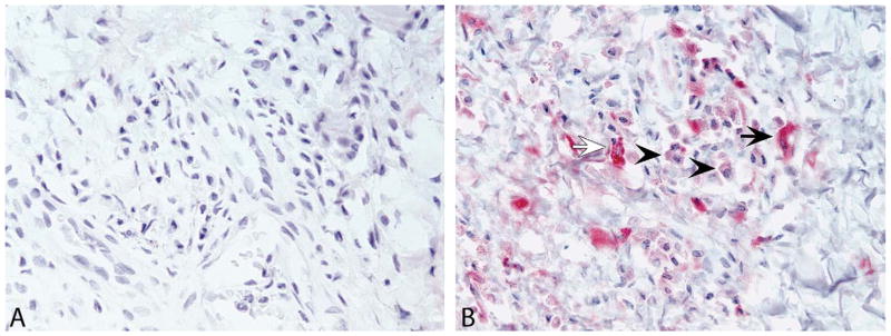
Immunohistochemistry for EBV antigens EBNA2 and LMP1 in sections of genital lesions from a Mauritian-origin cynomolgus macaque (Case No. 14). Representative images show negative immunolabeling for the Epstein-Barr virus (EBV) nuclear antigen EBNA2 (A) and moderate to strong cytoplasmic labeling for latent membrane protein 1 (LMP1; B) in low numbers of intralesional spindle-shaped stromal cells (arrows) and plasma cells (arrowheads). Images were taken at 40× objective magnification.