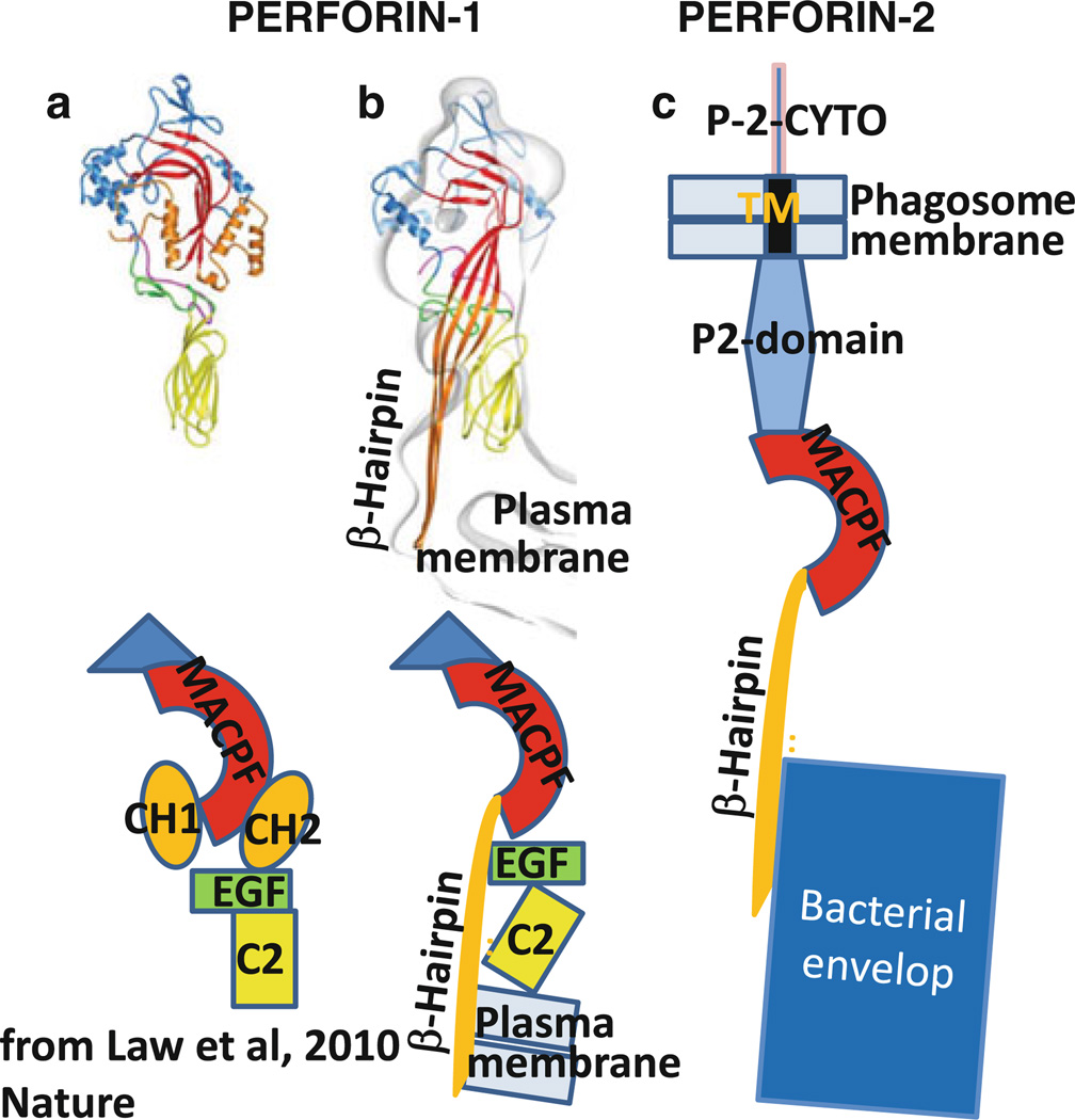
Fig. 13.
Model of perforin-2 refolding and killing bacterium based on crystal structure of perforin-1 (Law et al. 2010). a Crystal structure and schematic domain structure of perforin-1. b Refolded perforin-1 inserted into lipid bilayer (plasma membrane). c Refolded perforin-2 attacking bacteria inside phagosome. Note that perforin-2 remains tethered to the phagosome membrane via its transmembrane domain

