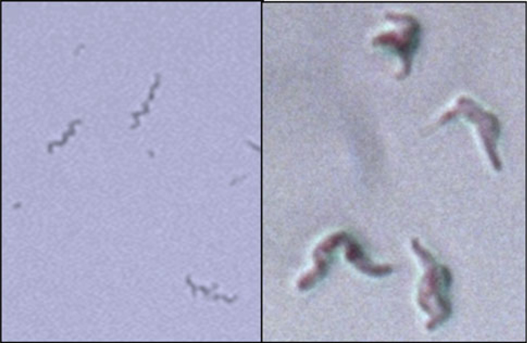
Fig. 9.
Left: M. smegmatis from fresh liquid culture. Right: M. smegmatis isolated from IFN-activated fibroblasts 5 h after infection and then plated on agar overnight. Note the intact but grotesquely swollen body of M. smegmatis. Addition of lysozyme does not affect the images on the left but disintegrates M. smegmatis on the right to small fragments and debris (not shown)

