
Figure 4.
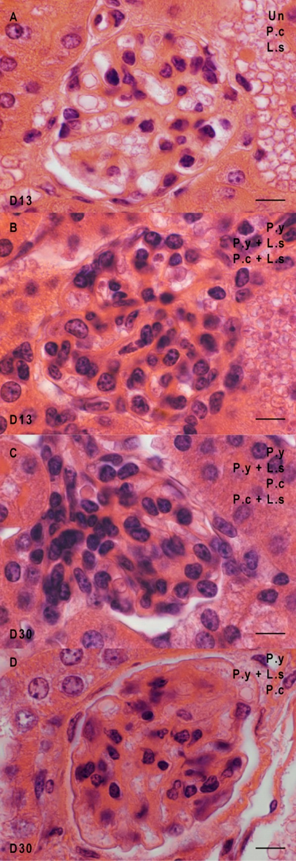
Glomerulonephritis in the kidneys of the mice at different times of infection. Glomeruli from uninfected mice (uninf.), mice infected by L. sigmodontis (L. s), mice infected by P. yoelii (P. y), mice infected by P. chabaudi (P. c), mice co-infected by P. y + L. s and mice co-infected by P. c + L. s were analysed at two time points, e.g., D13 and D30. (A) Normal glomeruli at day 13 post-infection. (B) Proliferation of the mesangial cells at day 13 in both co-infected groups and in P. y-infected mice. (C) Proliferation of the mesangial cells at day 30 in both plasmodial infected groups and in both co-infected groups. (D) Obliteration of the capillaries at day 30 in both plasmodial infected groups and in the mice co-infected with L. s and P. y. Scale bars represent 10 μm.