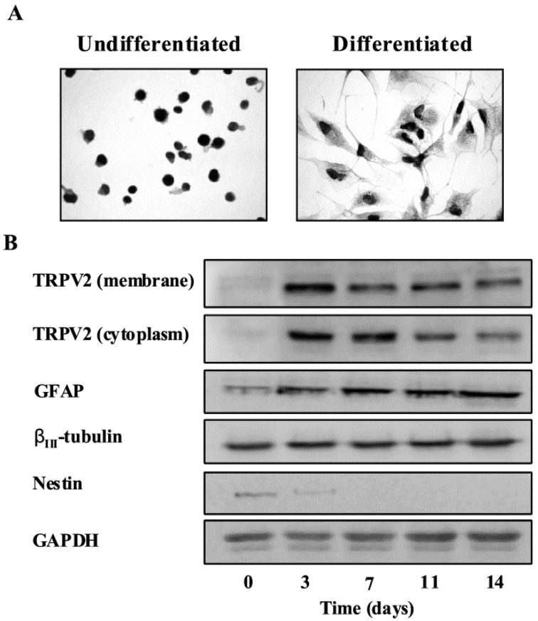
Figure 1.
TRPV2 protein expression during GSC differentiation. (A) TRPV2 protein expression in undifferentiated and differentiated GSCs was evaluated by immunocytochemistry analysis. GSCs were incubated with goat anti-TRPV2 antibody followed by the respective secondary antibody. The detection was performed by avidin-biotin complex peroxidase method and diaminobenzidine (DAB) as a chromogen. The result shown that a moderate reaction that stains all the cytoplasm occurs in differentiated GSCs compared to the undifferentiated GSCs. (B) Membrane and cytoplasm fractions from undifferentiated or differentiated GSCs were immunoblotted with anti-TRPV2 antibody. Total lysates were also immunoblotted with anti-GFAP, anti-βIII-tubulin and anti-nestin antibodies. GAPDH protein was used as loading control.
Adapted from 1 [54] Copyright John Wiley and Sons 2012. Permission obtained from copyright holder.

