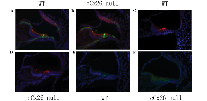
Figure 1.
Cellular structures of the organ of Corti at P0 were compared between (A, C and E) WT and (B, D and F) cCx26 null mice. Hair cell and supporting cell specific markers were immunolabeled. All cell nuclei were stained with DAPI (4′,6-Diamidino-2-Phenylindole). (A and B) Supporting cells (pillar cells) were marked with P75 antibody (green fluorescence). Red fluorescent labeling with phalloidin was used to visualize the cell borders. (C and D) Hair cells (including one inner hair cell and three outer hair cells) were labed with mysin6 (red fluorescene). (E and F) Supporting cells were marked with Prox1 antibody (green fluorescence). cCx26 null, conditional connexin 26 knockout mouse; WT, wild type mouse.

