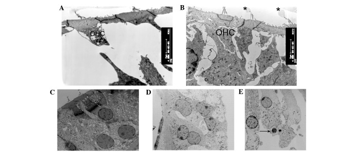
Figure 5.
Ultrastructural changes of the outer hair cells regions. (A and B) At postnatal day 10 (P10), the majority of outer hair cells appeared cuboid with enlarged cytoplasm in cCx26ko mice compared with wild-type mice (magnification, ×4,800; middle turn). (C and D) Following P18, outer hair cells became cuboid with unclear cell boundaries and little intercellular space (magnification, ×1,680; middle turn). (E) At P60, nuclei of outer hair cells showed pycknosis and cells were severely degenerated (magnification, ×3,800; basal turn). Arrows, degenerated cells; OHC, outer hair cells; cCx26ko, conditional connexin 26 knockout.

