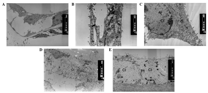
Figure 6.
Ultrastructural changes of Claudius cells. (A) At postnatal day 8 (P8), the Claudius cells of cCx26ko mice appeared normal (magnification, ×2,850; apical turn). At P10, (B) mitochondria of wild-type mice were dense and robust (magnification, ×8,400; apical turn) and (C) mitochondria of cCx26ko mice were enlarged (magnification, ×8,400; middle turn). Following P18, (D) Claudius cells degenerated and (E) cytoplasm became scarce and scattered (magnification, ×4,800; middle turn). Fewer mitochondria were observed in Claudius cells at P30 in cCx26ko mice (magnification, ×6,500; middle turn). Cl, Claudius cells; M, mitochondria; cCx26ko, conditional connexin 26 knockout.

