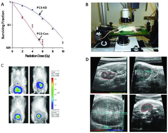
Figure 2.
(A) Clonogenic survival assay using a PC3-Con (DAB2IP proficient) PC3-KD (DAB2IP silenced) cell line. (B) Set up of ultrasound imaging station for tumor cell implantation and imaging. (C) BLI of four different Copenhagen rats demonstrating various stages of tumor growth. (D) Ultrasound imaging of prostate as well as proximal pelvic organs. Prostate tumors are outlined in red. Each panel represents an axial ultrasound image of an OT tumor in the prostate. The left upper section of this image is notable for diffuse calcification and necrosis. Tumors reached diameters as large as 2 cm before being euthanized.

