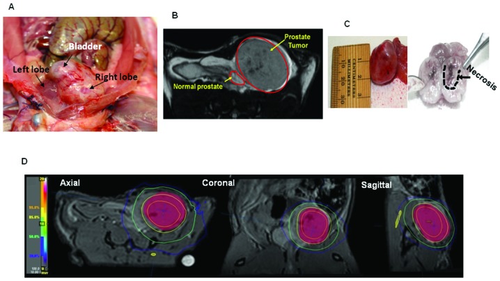
Figure 3.
(A) Digital image displaying OT tumors in the rat pelvis after euthanasia. No visible metastasis was observed to other structures within the perineum or peritoneum. (B) MRI provides a non-invasive method to track tumor growth. (C) Specimen tumor resected en bloc with representative sizing. The tumor, once dissected, displays large grossly visible areas of necrosis. (D) MRI was used to create radiation treatment plans for the rats for applying uniform dose.

