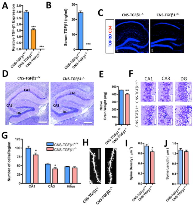
Figure 1. Characterization of CNS-TGF-β1-deficient mice.
(A) TGF-β1 mRNA (expression normalized to GAPDH) is undetectable in the hippocampus of CNS-TGFβ1−/− mice (n=3). (B) TGF-β1 protein is undetectable in the serum of CNS-TGFβ1−/− mice (n=3). (C) CD4+ T-cells are absent in the brains of both CNS-TGFβ1−/− and CNS-TGFβ1+/+ mice. (D) Photomicrographs of Nissl-stained brain sections from CNS-TGFβ1−/− and CNS-TGFβ1+/+ mice. The hippocampus of CNS-TGFβ1−/− animals appears normal in size and shape. Scale bar: 200 μm. (E) The brain of CNS-TGFβ1−/− mice is reduced in weight as compared to CNS-TGFβ1+/+ littermates. (F) The pyramidal-cell layer in TGF-β1-deficient animals appears condensed in distinct regions of the hippocampus. Scale bar: 10 μm. (G) 6–8 day-old CNS-TGFβ1−/− mice display significantly reduced numbers of neurons in the area CA1 and CA3 of the hippocampus. (H) Photomicrographs of dendritic spines on biolistically transfected pyramidal neurons in hippocampal slices from 21-day old CNS-TGFβ1−/− or CNS-TGFβ1+/+ mice. Scale bar: 5 μm. (I–J) Pyramidal cells from CNS-TGFβ1−/− display a reduced density of dendritic spines (8 days in vitro). [*p < 0.05; **p<0.01; ***p<0.001]

