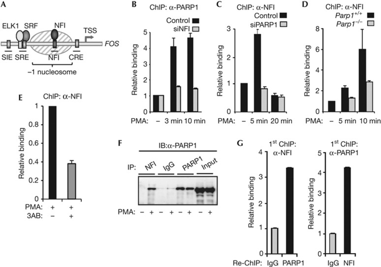
Figure 2.
PARP1 and NFI mutually aid each other’s association with the FOS promoter. (A) Schematic diagram of the FOS promoter showing the locations of cis-regulatory elements. The shaded oval indicates a positioned nucleosome. (B–E) ChIP showing the association of PARP1 (B) and NFI (C–E) with the FOS promoter in HeLa cells (B,C,E) or wild-type or parp1−/− mouse MEFs (D). Where indicated, HeLa cells were transfected with siRNAs against NFI, PARP1 or GAPDH; cells were serum-starved (−) or starved and treated with PMA for 10 min (+) or for times indicated. In (E), cells were treated with the PARP inhibitor 3AB for 30 min before PMA stimulation. (F) Immunoprecipitations were carried out using NFI, PARP1 or control IgG antibodies followed by immunoblotting (IB for PARP1). HeLa cells were serum-starved (−), or starved then stimulated with PMA for 10 min (+). (G) Re-ChIP analysis of NFI and PARP1 co-association with the FOS promoter. HeLa cells were starved and treated with PMA for 10 min. Data in (B), (C), (D) and (E) are presented as means±s.e.m. and are the average of at least two independent experiments performed in at least duplicate (n⩾4). Data in (G) are presented as means±s.d. and are representative of at least two independent experiments performed in triplicate (n=3). ChIP, chromatin immunoprecipitation; GAPDH, glyceraldehyde 3-phosphate dehydrogenase; IB, immunoblotting; IgG, immunoglobulin G; MEF, murine embryonic fibroblast; PMA, phorbol myristate acetate; PARP1, Poly [ADP-ribose] polymerase 1; siRNA, small interfering RNA; WT, wild type.

