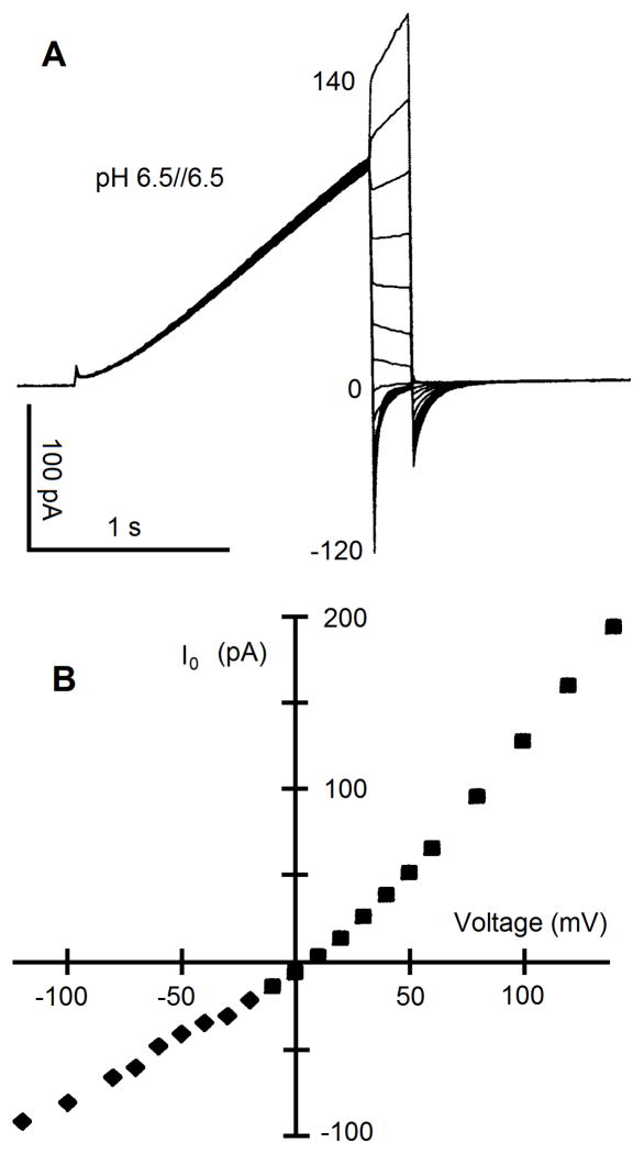
Fig. 7.
Measurement of the instantaneous current-voltage relationship of proton channels. In A, currents during repeated voltage pulses are superimposed. The first step, the “prepulse,” is to a voltage at which the gH is activated, and then the membrane is stepped to a range of voltages. Ideally, the same number of channels is open at the end of each prepulse, and therefore the current at the start of the test pulse (B) reflects any rectification of the open channel current. If one measured the current-voltage relationship of a single open channel, the result should have the same shape as that plotted in B. If any gating that occurs during the test pulse, typically channels closing at negative voltages, is so rapid that the initial current cannot be resolved, one can fit the decaying current, usually to an exponential decay function, and extrapolate back to the start of the pulse. From reference (90).

