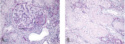
Figure 2.

Histopathology of oligomeganephronia. A) Virtually all glomeruli in the sample were at least twice normal size. This glomerulus contains segmental zones of perihilar glomerulosclerosis (arrows) and a voluminous juxtaglomerular apparatus. Fibrosis, tubular atrophy and chronic inflammation are evident in parts of the adjacent parenchyma. The scale bar represents 50 µm [Periodic Acid Schiff (PAS), x200]. B) As shown here, all intact tubules in the sample were hypertrophic. Diameters of the tubules were two to three times normal. The scale bar represents 50 µm (PAS, x200).
