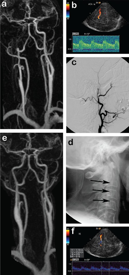
Figure 1.

A) Contrast enhanced magnetic resonance angiography (CE-MRA) shows occlusion of the right internal carotid artery. B) Axial transcranial color-coded sonography (TCCS) through the right temporal bone window demonstrating reversal of flow in the right anterior cerebral artery into the middle cerebral artery (both in red) with blood flow velocities within normal limits. C) Lateral DSA of the right common carotid artery showing stump of the inter-nal carotid artery which appears totally occluded. D) Late phase of the DSA series re-veals delayed antegrade filling of the ICA. E) Postoperative CE-MRA with regular filling of the right internal carotid artery. F) Postoperative TCCS with normal blood velocities in the right middle cerebral artery (in red) and normalization of flow in the anterior cerebral artery (now blue).
