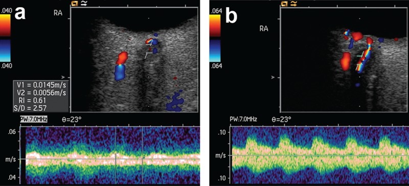
Figure 2.

A) High resolution orbital sonography with the doppler sample volume placed into the hypoechoic optical nerve with the residual low flow in the central retinal artery. B) Post-operatively orbital sonography demonstrates normal flow in the central retinal artery which can now be easily delineated together with its vein in red and blue.
