
Figure 2.
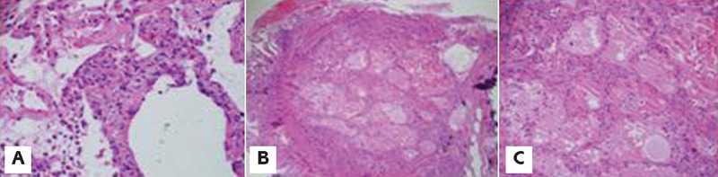
Photomicrograph of transbronchial lung biopsy. A) cellular interstitial pneumonitis with interstitial septal expansion by a mixed inflammatory infiltrate; B) and C) Prominent alveolar edema characterized by pale edema fluid within alveolar spaces (low and high power respectively).