
Figure 8.
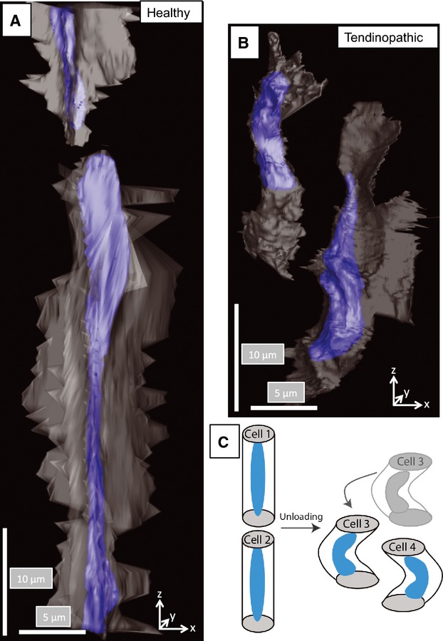
Buckling and slippage of cells in tendinopathic regions. (A) Three-dimensional reconstruction showing two cells in healthy tendon. The large cell in the center of the view is undergoing mitosis and is aligned parallel to the tendon long axis. Both cells are stacked one-on-top of the other. (B) Three-dimensional reconstruction showing two cells in tendinopathic tendon. The cells are aligned side-by-side, which was not observed in healthy tendon. (C) Schematic representation of cell slippage to explain the change in cell orientation from head-to-tail to side-by-side alignment.