Figure 2.

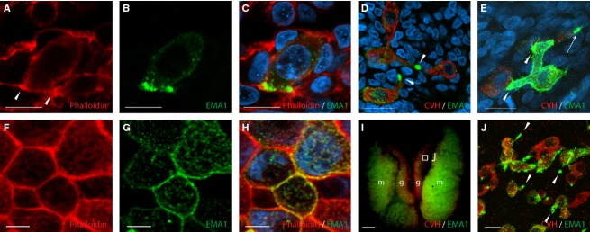
EMA1 epitope on PGCs is concentrated at sites of cell–cell contact. Whole-mount immunofluorescence of HH stage 20 embryos, demonstrating phalloidin staining of actin fibers (A) and EMA1 staining (B), co-localized to the elongated edges of a PGC, the contact sites with neighboring cells (C) or with other PGCs (D, E, arrowheads). Polarized EMA1 staining is also demonstrated for PGCs in the gonads of E7 embryos (I, J). (J) is an enlargement of the boxed area in (I). Epithelial cells examined at similar stages show non-polarized actin staining (F) and uniform EMA1 labeling at the cell surface (G, H). CVH, chicken vasa homolog; g, gonad; m, mesonephros. Scale bar: 10 μm (A–E); 5 μm (F–H); 1 mm (I); 50 μm (J).
