Figure 3.
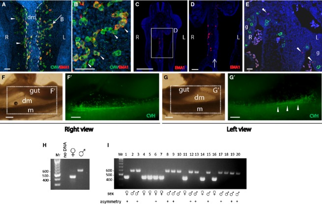
Left–right asymmetry in migration of PGCs through the dorsal mesentery. Immunofluorescence on whole-mount (A–B, F', G') and paraffin sections (C–E) of HH stages 20–22 embryos, using chicken vasa homolog (CVH) and EMA1 antibodies. (A) In HH stage 20 embryos, PGCs are present on the right but not left side of the mesentery. The PGCs on the left side are observed migrating toward the genital ridge. Cells were detected as clusters (arrowheads) or chains (arrow). (B) Field magnification of PGC-chain indicated by an arrow in (A). Examples for polarized EMA1 staining are indicated by arrowheads. (C, D) Coronal section of HH stage 20 embryo demonstrating PGCs on the right side of the mesentery. Area of condensed mesodermal cells on the left side of the mesentery is indicated by an arrow. (E) Co-labeling of EMA1 and pHistone-H3 demonstrating the asymmetrical distribution of PGCs vs. equal distribution of pHistone-H3 (arrowheads). (F–G') Right (F–F') and left (G–G') views of the mesentery of a HH stage 22 embryo immunostained for CVH (F',G'). PGCs can be seen on the right side of the mesentery (F'). A left view of the same area reveals only a faded signal of PGCs from the right side (G'), while most PGCs have reached the genital ridge (G', arrowheads). (H) Sex determination using PCR. Negative control (no DNA) and positive controls (female and male) are shown. (I) PCR results of 20 representative DNA samples, isolated from chicken embryos. The sex of each embryo and the presence of asymmetry in migration are indicated. dm, dorsal mesentery; g, gonad; L, left; m, mesonephros; R, right. Scale bar: 50 μm (A, B); 80 μm (C, D); 1 mm (E, E', F, F').

