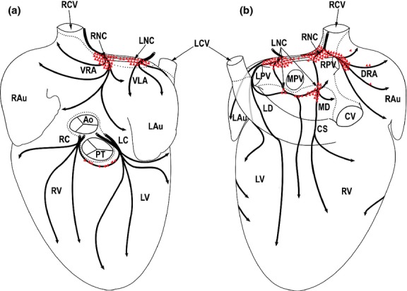
Figure 1.

Drawings summarizing the morphological pattern of distinct epicardial nerve subplexuses from 11 rabbit hearts as seen from the ventral (a) and dorsal (b) views of the pressure-distended heart stained histochemically for acetylcholinesterase (AChE). The clusters of intrinsic cardiac neurons (ICNs) (drawn in red) were outlined from the whole-mount stained histochemically for AChE. Dotted lines demarcate limits of the heart hilum. Black arched arrows indicate the course of nerve subplexuses on the rabbit heart surface. Red polygonal triangled areas indicate the location of neuronal clusters and epicardial ganglia. Ao, ascending aorta; CS, coronary sinus; CV, caudal vein; DRA, dorsal right atrial subplexus; ICNs, intrinsic cardiac neurons; LAu, left auricle; LC, left coronary subplexus; LCV, left cranial vein; LD, left dorsal subplexus; LNC, left neuronal cluster; LPV, left pulmonary vein; LV, left ventricle; MD, middle dorsal subplexus; MPV, middle pulmonary vein; PT, pulmonary trunk; RAu, right auricle; RC, right coronary subplexus; RCV, right cranial vein (superior caval vein); RNC, right neuronal cluster; RPV, right pulmonary vein; RV, right ventricle; VLA, ventral left atrial subplexus; VRA, ventral right atrial subplexus.
