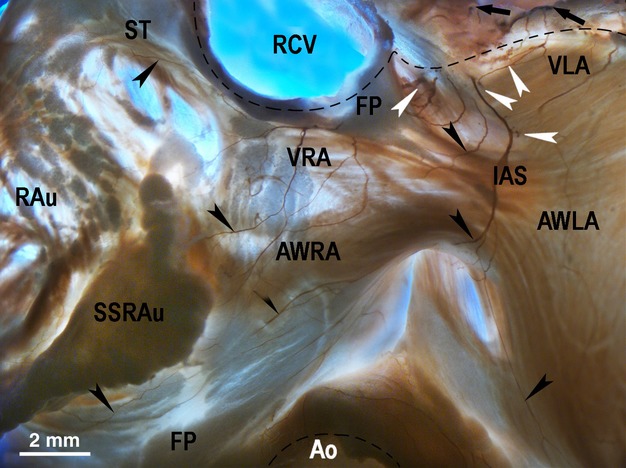
Figure 3.

Macrophotograph demonstrating the location and the morphological pattern of the ventral right (VRA) and left (VLA) atrial nerve subplexuses in the rabbit heart stained histochemically for acetylcholinesterase. Black arrows point to the access of extrinsic cardiac nerves into the heart through the venous part of the heart hilum (at the bifurcation of the pulmonary trunk). Black arrowheads point to some subplexal nerves extending to the innervation regions. White arrowheads point to the epicardial ganglia on the anterior wall of the root of the right cranial veins. Dashed line demarcates the heart hilum. Ao, aorta; AWLA, anterior wall of left atrium; AWRA, anterior wall of right atrium; IAS, projection of the interatrial septum; RAu, right auricle; RCV, right cranial vein; SSRAu, superior surface of right auricle; ST, sulcus terminalis; VLA, ventral left atrial subplexus; VRA, ventral right atrial subplexus.
