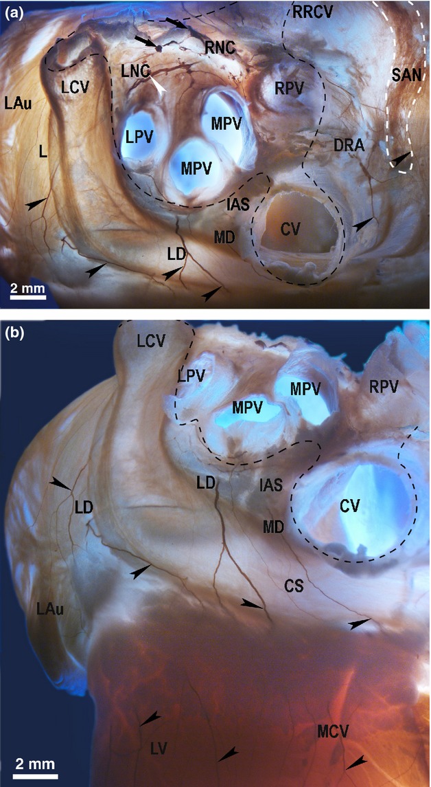
Figure 4.

The location of the left dorsal (LD) and the dorsal right atrial (DRA) neural subplexuses in the rabbit heart stained histochemically for acetylcholinesterase. A dorso-caudal view (a) and a caudal view (b). Black arrows point to the access of extrinsic cardiac nerves into the heart through the venous part of the heart hilum (at the bifurcation of the pulmonary trunk). White arrowhead indicates the ganglion of the left neuronal cluster (LNC). Black arrowheads point to some epicardial nerves of the LD, MD and DRA subplexuses extending to the innervation regions. Dashed line demarcates the venous part of the heart hilum. CS, coronary sinus; CV, orifice of caudal vein (inferior caval vein); DRA, dorsal right atrial subplexus; IAS, projection of the interatrial septum; LAu, left auricle; LCV, left cranial vein; LD, left dorsal subplexus; LNC, left neuronal cluster; LPV, left pulmonary vein; LV, left ventricle; MCV, middle cardiac vein; MD, middle dorsal nerve subplexus; MPV, middle pulmonary vein; RNC, right neuronal cluster; RPV, right pulmonary vein; RRCV, root of the right cranial vein (superior caval vein); SAN, region of sinus node (outlined by dashed white line).
