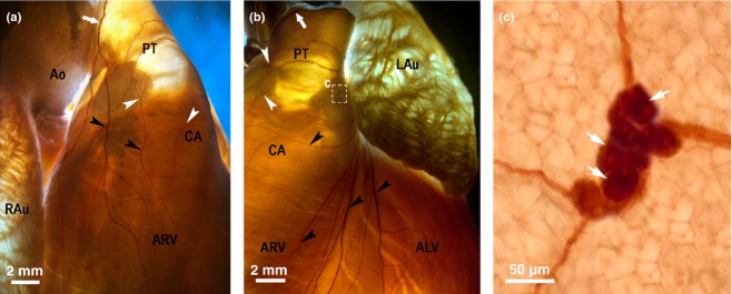
Figure 5.

Macrophotographs of the right (a) and the left (b) coronary subplexuses (RC and LC) in the rabbit heart stained histochemically for acetylcholinesterase. White arrows indicate the access of extrinsic cardiac nerves into the heart through the arterial part of the heart hilum. The boxed area ‘c’ in the panel (b) is enlarged as the panel (c). Black arrowheads point to some subplexal nerves extending to the innervation regions, the white ones point to some small epicardial ganglia on the conus arteriosus (CA) and the root of pulmonary trunk (PT). ALV, anterior wall of the left ventricle; Ao, root of the aorta; ARV, anterior wall of the right ventricle; LAu, left auricle; RAu, right auricle.
