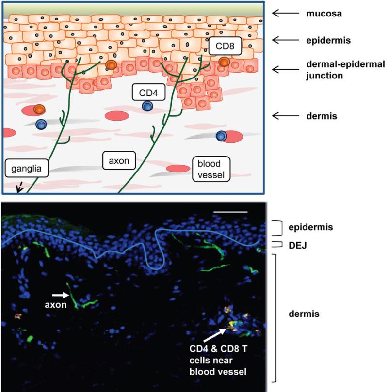
Figure 2. Schematic and histologic representation of healthy genital mucosa.
Axons arrive from the dorsal root ganglia and terminate at the dermal-epidermal junction or within the mid-layer of the epidermis. HSV-2 is released from axonal termini across the neuron-epithelial gap, and can enter epithelial cells where virus is amplified and rapidly spreads to contiguous skin cells. Low numbers of CD4+ and CD8+ T cells are present in the dermis and dermal-epidermal junction after entering the genital skin through blood vessels. The white line denotes the dermal-epidermal junction.

