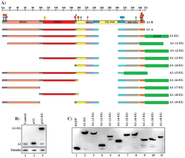
Fig. 1.
(A) Schematic representation of hnRNP A1 isoforms (A1-A, A1B) and deletion clones map. The protein RBDs and other key structural and functional features are indicated, M9 (nuclear localization sequence), F (F-peptide), PrLD (prion like domain). The location of residues modified by acetylation (A), phosphorylation (P) and SUMOylation (S) is indicated. The sequences present in the major deletion clones are indicated. (B) Expression of hnRNP A1 in control HEK-293 cells or transfected with the hnRNP A1-A expression clone (pA1) or the plasmid expressing the hnRNP A1-EGFP fusion protein. Proteins are detected with the anti-hnRNP A1 antibody. (C) Expression of the hnRNP A1–EGFP fusion proteins in HEK-29 cells. Proteins are detected with the anti-GFP antibody.

