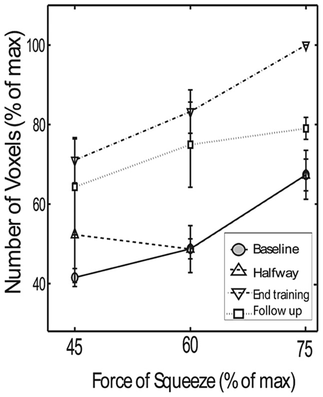
Figure 6.

Number of activated voxels in the contralateral SMC as a function of squeezing force in chronic stroke patients. Online mapping was performed at four time-points: baseline (solid line); halfway through training (lower dashed line); at the end of the training (upper dashed line) and 4 weeks after training (dotted line). Note the persistence of increased cortical activation observed during and after the training period.
