
Figure 1.
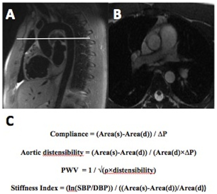
A–B. Scout cardiovascular magnetic resonance image of thoracic aorta demonstrating the planning of a transverse section through proximal descending aorta at the level of the right pulmonary artery. C. Arterial stiffness equations. Area(s) = systolic area, area(d) = diastolic area, ΔP = SBP-DBP, ρ = blood density (1059 kg.m−3).