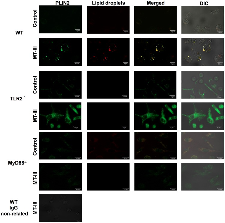
Figure 4. TLR2 and MyD88 are essential for MT-III-induced perilipin-2 (PLIN2) subcellular distribution.
Wild Type (WT), TLR2−/− or MyD88−/− peritoneal macrophages were incubated with RPMI (control) or MT-III (0.4 μM) for 6 h and labeled for both lipid droplets (fluorescent Nile red) and anti-PLIN2 (FITC-conjugated immunocomplex). Merged image shows localization of PLIN2 to lipid droplets in WT macrophages. Cell nuclei were stained with propidium iodide. IgG control was included and showed negative stain. The micrographs are representative of at least three samples/experimental group.

