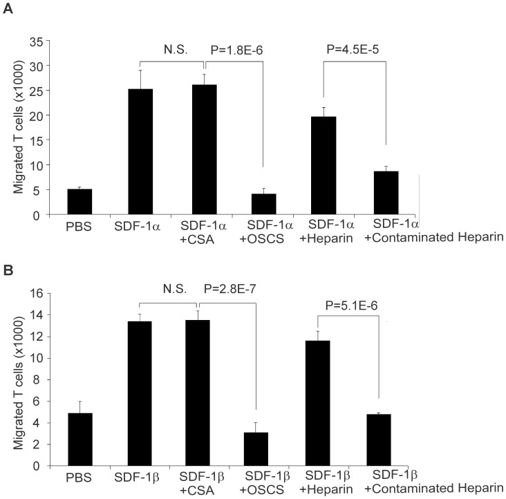
Figure 3. OSCS inhibits SDF-1-mediated T cell chemotaxis.
PHA and IL-2-induced human T cell blasts were added at 105/100 μL per well to the upper chambers of 24-well transwell plates. 1 mL serum-free culture medium with 100 ng recombinant SDF-1α (A) or SDF-1β (B), alone or in the presence of 25 μg/ml of CSA, OSCS, heparin or a heparin lot contaminated with OSCS were added to the lower chambers as described in the Materials and Methods section. After two hours of incubation at 37°C in a CO2 incubator, the cells that migrated to the lower chambers were counted by flow cytometry using BD Trucount tubes. P values of one-way ANOVA test among groups were less than 0.0001 for both (A) and (B) supporting the hypothesis that the population means are not all equal. P values based on Student’s t test of the differences between SDF-1 with CSA and SDF-1 with OSCS and between SDF-1 with heparin and SDF-1 with contaminated heparin are shown. There is no significant difference between SDF-1 and SDF-1 with CSA. The data are the averages of triplicates and the experiment has been repeated with cells from at least three different donors.

