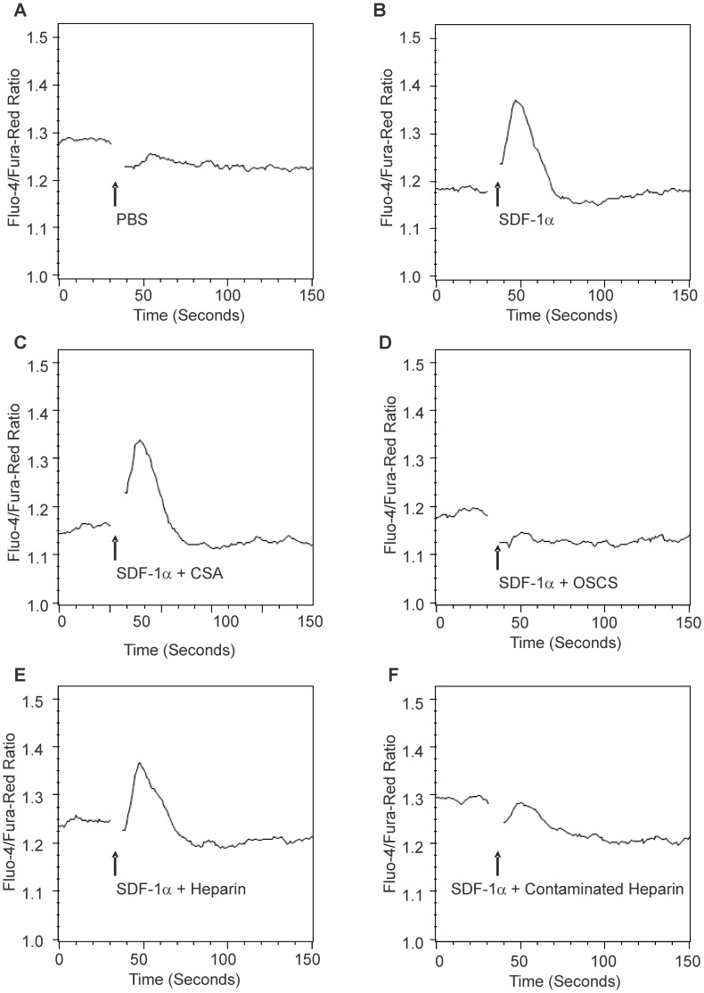
Figure 4. OSCS inhibits SDF-mediated T cell calcium mobilization.
PHA and IL-2 activated human T cell blasts were loaded with fluo-4 and fura red dyes as described in the Materials and Methods section. The T cell response to SDF-1α was detected using a LSRII flow cytometer and expressed as the fluorescence ratio of fluo-4/fura-red dyes. (A) The injection of PBS did not cause a calcium influx in T cells; (B) 100 nM SDF-1α induced a strong calcium influx in T cells; (C) SDF-1α in the presence of CSA induced a strong calcium influx in T cells; (D) SDF-1α in the presence of OSCS did not cause a calcium influx in T cells; (E) SDF-1α in the presence of heparin induced a calcium influx in T cells; (F) SDF-1α in the presence of heparin contaminated with OSCS did not induce a calcium influx in T cells. The data are representative of experiments with at least three independent donors.

