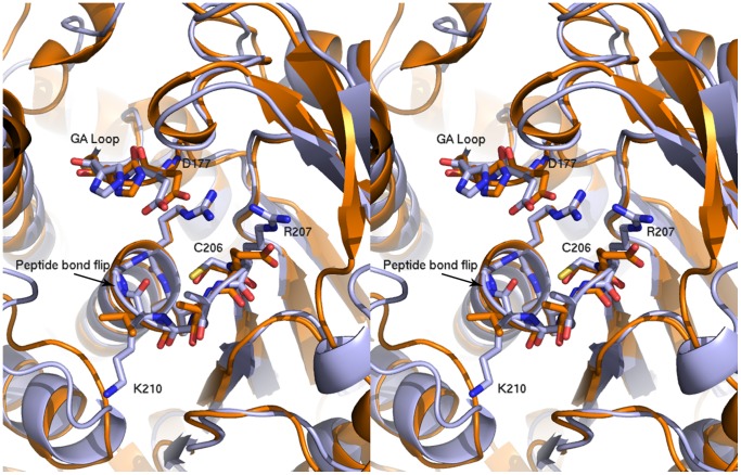
Figure 5. Comparison of the structure of the catalytic site in Bd1204 (Light blue) and PhyAsr (Orange).
The position of the catalytic residues, the phosphate binding loop (P-loop) and the general acid loop (GA loop) are conserved in both proteins. Basic residues and peptide bond flip observed in are labeled and shown as sticks. The protein is oriented the same as that shown in Figure 4.

