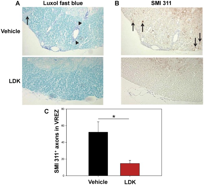
Figure 4. LDK provides axonal preservation during the peak of EAE-relapse.
Spinal cord sections obtained on Day 43, the peak of EAE-relapse, from vehicle-treated and LDK-treated SJL/J mice sensitized with PLP139–151 were stained with Luxol fast blue and SMI 311. (A) Representative Luxol fast blue stained spinal cord sections showed perivascular cuffing (arrowheads) and demyelination (arrow) in the vehicle-treated mouse in comparison to the LDK-treated mouse. (B) Representative staining of non-phosphorylated neurofilaments using SMI 311 on consecutive tissue sections showed staining in the white matter of the vehicle-treated mouse (arrows), which is indicative of axonal damage. However, SMI 311 is also able to bind to healthy dendrites and nucleated cells in the gray matter. (C) Quantification of SMI 311 positive axons in the white matter of the ventral root exit zone (VREZ) of vehicle-treated and LDK-treated mice at the peak of disease relapse. Results represent the mean+SEM for groups of 3 mice with at least 6 tissue slices per mouse scored by Image Pro-Plus. *P<0.05, Student’s paired t-test.

