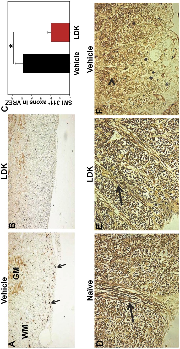
Figure 5. LDK prevents axonal damage in EAE-relapsed mice.
Spinal cord sections obtained on Day 56 from vehicle-treated and LDK-treated SJL/J mice sensitized with PLP139–151 were stained with SMI 311 and silver stain. (A) Representative spinal cord section of vehicle-treated mouse stained with SMI 311, indicated by the brown dots (arrows). Gray matter (GM) and white matter (WM). (B) Representative spinal cord section of LDK-treated mouse stained with SMI 311. (C) Quantification of SMI 311 positive axons in the white matter of the VREZ. Data is representative of the mean+SEM for groups of 7 mice with at least 5 tissue slices per mouse scored by Image Pro-Plus. *P<0.05, Student’s paired t-test. (D–F) Representative silver-stained spinal cord tissue sections of Naïve (D) SJL/J mice, used as a control, and SJL/J mice sensitized with PLP139–151 peptide and treated with either LDK (E) or vehicle (F). Intact axons (arrow) and blebs (arrowhead) are indicated. Images are 60× magnification of VREZ.

