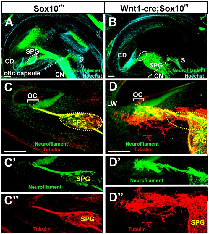
Figure 7. Immunochemistry of a coronally sectioned ear shows differences in spiral ganglion cell distribution and projection.
Spiral ganglion neurons (SGN) are inside the otic capsule in the control ear (A) but have migrated to the center of the spiral and partially outside the ear in the Sox10 mutant (B, dotted line). The dotted circle in (A) indicates spiral ganglion location and is shown in (B) to indicate the position of Rosenthal’s canal. Most spiral ganglion neurons are tubulin positive and few are neurofilament positive in control (C, C’, C”) and Sox10 mutant mice (D, D’, D”). However, while all labeled fibers run together from spiral ganglion neurons to innervate hair cells in control mice (C, C’ C”), many tubulin labeled afferent fibers of Sox10 mutant mice bypass the organ of Corti (D, D’, D”). Abbreviations: OC, organ of Corti; HC, hair cells; LW, lateral wall. Bar indicates 100 μm.

