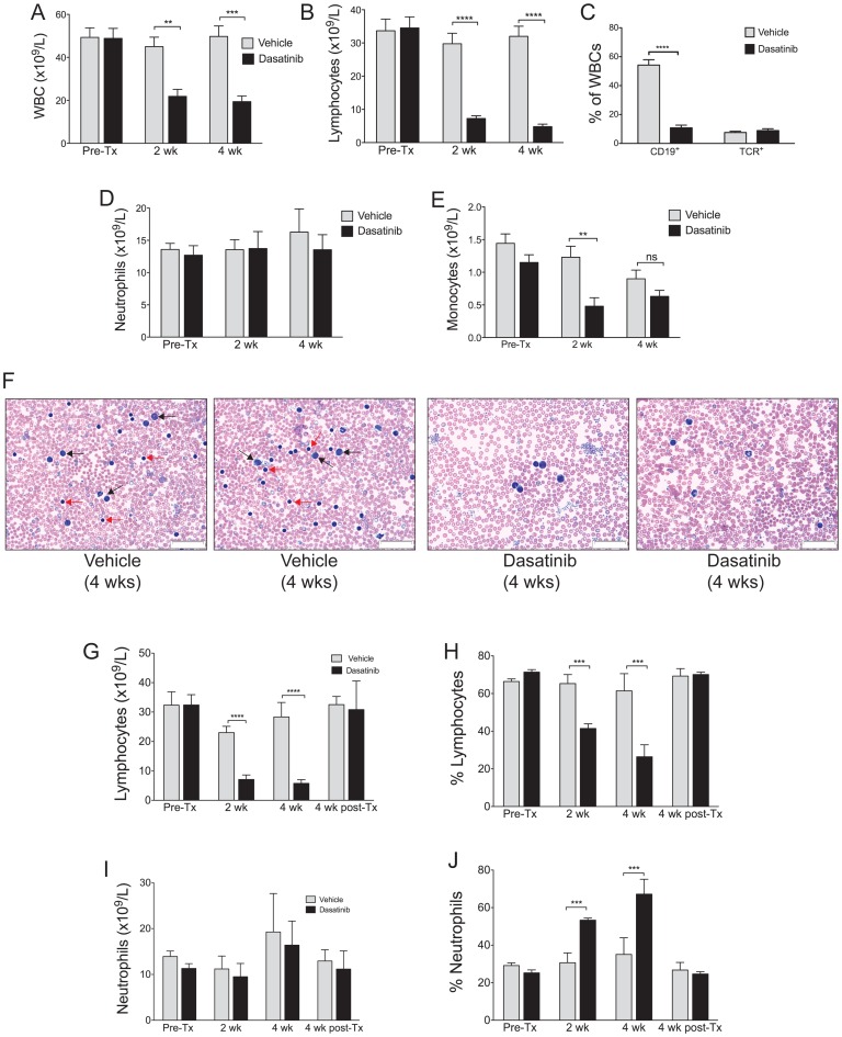
Figure 2. High dose dasatinib treatment results in a significant reduction of B lymphocytes in the blood.
c-Cbl RING finger mutant mice aged 8–9 months were dosed daily with 30 mg/kg (am) +50 mg/kg (pm) of dasatinib or vehicle, and bled before treatment (Pre-Tx), and after 2 and 4 weeks of treatment. Differential blood counts from 9 vehicle and 7 dasatinib treated mice were determined by Hemavet analysis. Shown are (A) total WBC numbers and (B) lymphocyte numbers. (C) WBCs were analyzed by flow cytometry to determine the percentage of B-lineage cells, by anti-CD19 staining, and the percentage of T cells, by anti-T cell receptor staining. (D) Numbers of neutrophils and (E) monocytes. (F) Blood films from 2 vehicle and 2 dasatinib treated mice following 4 weeks of dosing illustrate the loss of lymphocytes in the dasatinib-treated mice. Lymphocytes are indicated by red arrows, and myeloid cells by black arrows, in the blood films from the two vehicle treated mice. The images were acquired at room temperature using an Olympus BX51 microscope with a 60×/0.09 objective and photographed with a SIS 3VCU Olympus digital camera. Scale bar = 50 μm. Lymphocyte numbers (G) and percentages (H) in the blood return to pre-treatment levels 4 weeks after ceasing dasatinib treatment. Neutrophil numbers remain unaltered (I) but the high percentages (J) returned to normal 4 weeks after dasatinib dosing ceases. Mice were bled from the tail vein before treatment (Pre-Tx), after 2 and 4 weeks of treatment, and following 4 weeks without treatment (i.e. post-Tx). Numbers and percentages were determined by Hemavet differential counting, and are from 4 vehicle and 3 dasatinib treated mice. The results are expressed as means ± standard errors. **P<0.01, ***P<0.001, ****P<0.0001 using unpaired Student’s t test.

