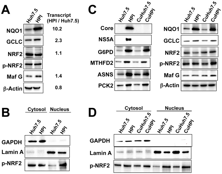
Figure 8. Expression of Nrf2, Maf G, and Nrf2-targert genes.
(A) Immunoblot analyses of the proteins related to antioxidation and detoxification, Nrf2, phosphorylated Nrf2 (p-Nrf2), and Maf G for Huh7.5 and HPI cells. Fold expression of transcript (HPI/Huh7.5) in corresponding genes was shown in the right according to the expression array data. (B) Immunoblot analysis of p-Nrf2 in cytosol and nucleus fractions from the cells used in (A). GAPDH and Lamin A were used as a marker protein for cytosol and nucleus, respectively. (C) Immunoblot analyses of the HCV core and NS5A proteins, Nrf2-trarget genes, Nrf2, p-Nrf2 and Maf G for Huh7.5 and HPI cells, CuHuh7.5 cells (Huh7.5 cells simply treated with cyclosporine) and CuHPI cells (HPI cells, from which HCV was eliminated with cyclosporine). (D) Immunoblot analysis of p-Nrf2 in their cytosol and nucleus fractions from the cells used in (C).

