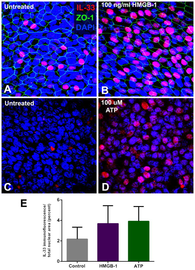
Figure 3.
Stimulation of sinonasal epithelial cell expression of IL-33 with HMGB-1 and ATP. Sinonasal epithelial cells grown at the air-liquid interface were exposed to the damage-associated molecules, HMGB-1 (100 ng/ml) for 24 hours, or ATP (100 μM) for 8 hours. A marked increase in nuclear expression of IL-33 protein was observed in recalcitrant CRSwNP subjects, although the mean increase in nuclear immunofluoresence did not achieve statistical significance for the group as a whole. (A/B: pre- and post-HMGB-1 exposure; C/D: pre- and post-ATP exposure). E: Graphical representation of increase in mean IL-33 immunostaining following exposure to HMGB-1 and ATP, across all subjects (p=ns).

