
Figure 2.
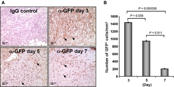
Nanofiber-expanded CD34+ cells were recruited to the wound bed. (A) Green fluorescence protein (GFP) was overexpressed on nanofiber-expanded CD34+ cells using Amaxa nucleoporation system, were assessed for homing to the wound bed at various time-points using α-GFP Ab by immunohistochemical methods. (B) Quantitative values of GFP+ cells were presented graphically. GFP+ cells were quantified by counting the cells in randomly chosen eight high-power microscopic fields within the wound-edge sections (n = 3) obtained from various time-points (day 3, 5 and 7). Data are presented as mean ± SEM.