Figure 6.

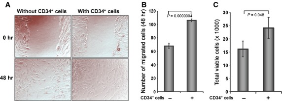
CD34+ cells enhance dermal fibroblast cell number and migration. (A) Cell migration assay was performed in two-chambered 24-well plate. Confluent dermal fibroblast was grown in lower chamber and monolayer was scratched with a p200 pipette tip. CD34+ cell containing inserts or without cell inserts (as a control) were placed on the top of well with scratched fibroblast cells. Images for each well were captured immediately after scratch and after 48 hrs of scratching. (B) Number of migrated fibroblast cells were counted in the presence or absence of CD34+ cells and presented graphically. (C) Number of fibroblast cells was counted from the bottom chamber of trans well (24-well) plate after 48 hrs of co-cultured with CD34+ cells or media. Non-adherent cells were removed by washing from the lower chamber before harvesting fibroblasts for analysis. Results are shown as mean ± SEM (n = 6) within a representative of three independent experiments.
