Fig. 2.
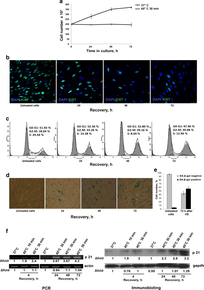
Heat shock (45 °C, 30 min) induced MSC premature senescence. a Growth curves of heated and unheated cells. MSCs exposed to heat shock stop division. b Immunofluorescence assay of cell proliferation with anti-Ki-67antibodies. Only single cells are Ki-67-positive 72 h after heat shock. c FACS assay of cell cycle distribution. MSCs that were heated at 45 °C for 30 min and returned to the normal culture conditions exhibited cell cycle arrest in G2/M phases. d SA-β-X-gal staining during cell recovery from sublethal HS. The number of X-gal-positive cell increased. e Quantitative assay of X-gal-positive cells. f Expression of cyclin-dependent kinase inhibitor p21 in MSCs exposed to HS. p21 expression is increased along with the augment in X-gal-positive cells (d)

