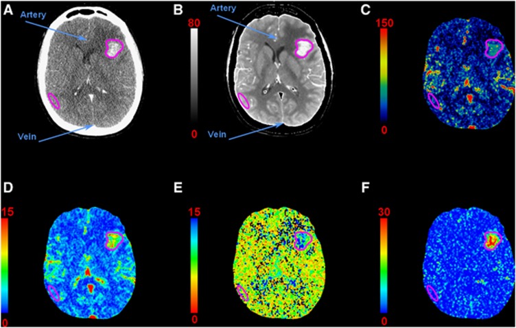
Figure 2.
Computed tomography (CT)-perfusion maps of a patient with a single metastasis in the left hemisphere: (A) contrast-enhanced CT, (B) temporal averaged image (HU), (C) cerebral blood flow (CBF) map (mL/min per 100 g), (D) cerebral blood volume (CBV) map (mL/100 g), (E) mean transit time (MTT) map (seconds), (F) permeability surface (PS) area product map (mL/min per 100 g). Regions of interest (ROIs) are drawn in the artery, vein, metastasis, and gray matter (GM) for perfusion measurements. Each pixel of a given map is expressed in the corresponding perfusion parameter unit. HU, Hounsfield units.

