Figure 2.

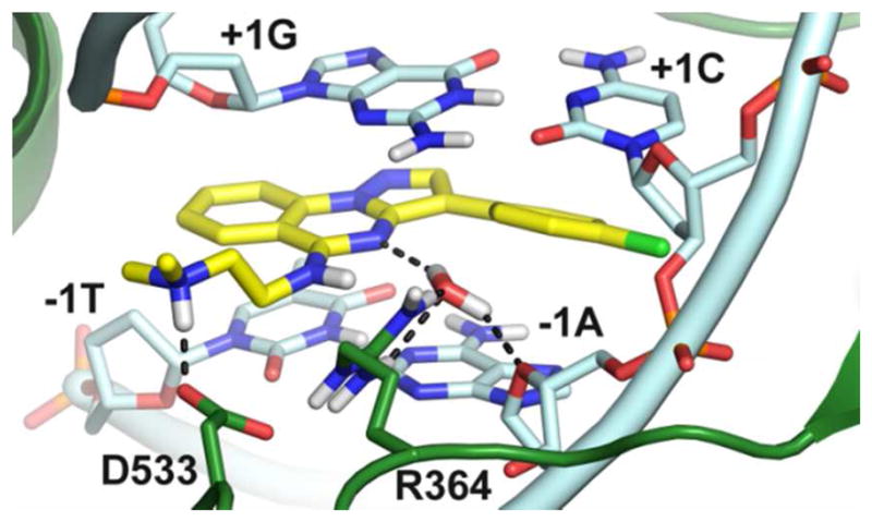
Binding mode of 26 (yellow sticks) at the Top1 (green cartoons) DNA (cyan cartoons) cleavage site. Residues important for ligand binding are highlighted as sticks. H–bonds are dashed black lines.

Binding mode of 26 (yellow sticks) at the Top1 (green cartoons) DNA (cyan cartoons) cleavage site. Residues important for ligand binding are highlighted as sticks. H–bonds are dashed black lines.