Fig. 5.
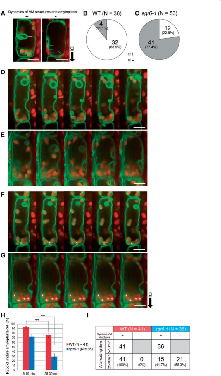
Dynamic vacuolar membrane (VM) structures and amyloplast dynamics in endodermal cells of cut stems. The endodermal cells of the transgenic plant expressing green fluorescent protein–tonoplast intrinsic protein-γ (GFP–γ-TIP), a lytic vacuole marker, under control of the SCARECROW (SCR) promoter, were observed using a vertical microscope. Green, GFP fluorescence; red, autofluorescence of Chl from plastids. (A) Endodermal cells that show or do not show invaginated VM structures and amyloplast dynamics at a time point within 5–10 min after cutting the stem. (B, C) Number of endodermal cells that show or do not show invaginated VM structures at a time point within 5–10 min after cutting the stem of the wild type (B) and sgr6-1 (C). At least eight stem segments of each genotype were observed. (D, E) Confocal images of wild-type (D) and sgr6-1 (E) endodermal cells observed for 2 min within 5–10 min after cutting the stem were aligned at approximately 30 s intervals. (F, G) Confocal images of wild-type (F) and sgr6-1 (G) endodermal cells observed for 2 min within 25–30 min after cutting the stem were aligned at approximately 30 s intervals. (H) The ratio of amyloplasts that moved along the x- or y-axis or that rotated during the 2 min observation. Endodermal cells that harbor invaginated VM structures at the starting point of observation within 5–10 min after cutting the stem were analyzed. At least 16 stem segments of each genotype were observed. Statistical differences between 5–10 min and 25–30 min were detected using Student’s t-test (**P < 0.01). Bars represent the SEs. (I) Number of endodermal cells that maintained invaginated VM structures during the 30 min observation. ‘g’ and a black arrow indicate the direction of gravity. Scale bars = 10 µm.

