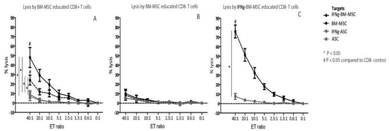
Figure 3ABC.
A: Dose dependent lysis of BM-MSC by BM-MSC educated CD8+ T cells (black dashed line). Lysis increased when BM-MSC targets were IFNγ-stimulated (black solid lines) and there was no lysis of the mismatched ASC (grey lines) indicating HLA-class I specific lysis. Mean ± SEM, n=5.
B: The BM-MSC educated CD8− T cells were not capable of lysing any of the target populations. Mean ± SEM, n=5.
C: When CD8+ T cells were educated with IFNγ-stimulated BM-MSC, lysis of IFNγ-stimulated BM-MSC targets increased (black solid line). Lysis was HLA-class I specific as IFNγ-BM-MSC educated CD8+ T cellsdid not lyse the mismatched ASC (grey lines). Mean ± SEM, n=5.

