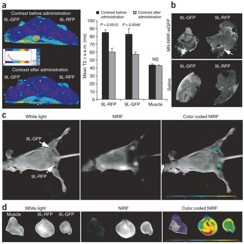
Figure 6.
Theranostic approach using dextran coated iron oxide nanoparticles (a) In vivo MRI was performed on mice bearing bilateral green fluorescent protein (9L-GFP) and red fluorescent protein (9L-RFP) tumors before and 24 h after nanoparticles administration. After injection of the probe, there was a significant drop in T2 relaxivity associated with the tumors. Note that T2 relaxation times of muscle tissue remained unchanged. (b) Ex vivo high-resolution MRIs of excised tumors (78 m isotropic). Distinct foci of signal loss (arrows), reflecting probe accumulation, were easily identifiable in tumors derived from mice injected with the probe but not from saline-injected controls. (c) In vivo NIR optical imaging of the same mice as in a produced a high-intensity NIR signal associated with the tumors. This confirmed the delivery of the nanoparticles probe to these tissues. (d) Ex vivo NIR optical imaging demonstrated a significantly higher fluorescence in tumors than in muscle tissue (P = 0.0058). Reproduced with permission from Figure 3 of ref. 60, Macmilan Publishers Ltd: Nature Medicine, © (2007).

