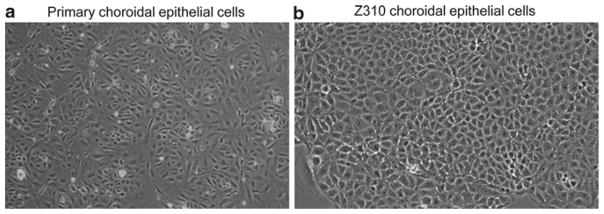
Fig. 2.

Morphology of choroidal epithelial cells in culture. (a) Primary culture of choroidal epithelial cells after 5 days in culture (10×). Note the con fluent layer of cells with a predominant polygonal cell type. The choroid plexus tissue was obtained from 6-week-old Sprague-Dawley rats. (b) Immortalized Z310 choroidal epithelial cells in culture (20×). Passage 86.
