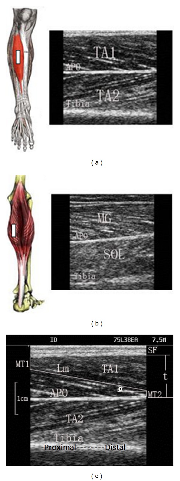
Figure 1.

Probe positions on the measured muscles and typical ultrasound images for measurement on (a) TA and (b) MG. (c) Demonstration of the labels for muscle parameters. The bright fringe in the lower region of the image shows the muscle-tibia boundary. Aponeurosis (APO) is the boundary between the superficial and deep layer of TA. SF is subcutaneous fat. L m is the visualized part of the entire muscle fascicle length and can be measured directly; MT1 and MT2 are the distance of the fiber proximal end point to the superficial aponeurosis and the distance of the fiber distal end to the bone, respectively; α is the pennation angle; TA1 is the superficial layer of the TA; and TA2 is the deep layer of TA.
